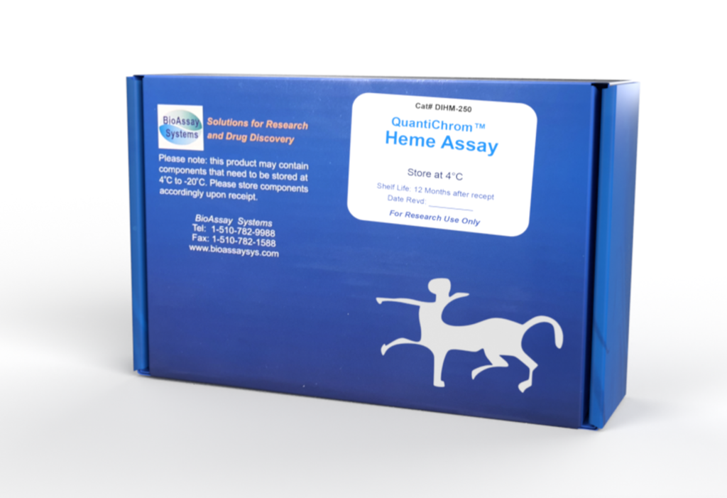
DESCRIPTION
Heme is one important member of the porphyrin family. It is synthesized in both mitochondria and cytoplasm, and is a key prosthetic group for various essential proteins such as hemoglobin, cytochromes, catalases and peroxidases. Heme determination is widely practiced by researchers of various blood diseases.
Simple, direct and automation-ready procedures for measuring heme concentration are becoming popular in Research and Drug Discovery. BioAssay Systems' QuantiChromTM Heme Assay Kit is based on an improved aqueous alkaline solution method, in which the heme is converted into a uniform colored form. The intensity of color, measured at 400 nm, is directly proportional to the heme concentration in the sample. The optimized formulation substantially reduces interference by substances in the raw samples and exhibits high sensitivity.
APPLICATIONS
Direct Assays: total heme in blood, serum, plasma, urine, hemecarrying enzymes. Pharmacology: effects of drugs on heme metabolism.
Drug Discovery: HTS for drugs that modulate heme levels.
KEY FEATURES
Sensitive and accurate. Linear detection range 0.6 – 125 µM heme in 96-well plate assay. Simple and high-throughput. The “mix-and-read” procedure involves addition of a single working reagent and reading the optical density. Can be readily automated as a high-throughput assay in 96-well plates for thousands of samples per day.
Safety. Reagents are non-toxic.
Versatility. Assays can be executed in 96-well plate or cuvette.
KIT CONTENTS (250 tests in 96-well plates)
Reagent: 50 mL Calibrator:
10 mL (equivalent to 62.5 µM heme)
Storage conditions. The kit is shipped room temperature. Store reagent and Calibrator at 4°C. Shelf life: 12 months after receipt.
Precautions: reagents are for research use only. Normal precautions for laboratory reagents should be exercised while using the reagents. Please refer to Material Safety Data Sheet for detailed information.
PROCEDURES
Procedure using 96-well plate:
1. Blank and Calibrator. Pipette 50 µL water (Blank) and 50 µL Calibrator into wells of a clear bottom 96-well plate. Transfer 200 µL water into the blank and Calibrator wells. The diluted calibrator is equivalent to 62.5 µM heme.
2. Samples. Serum and plasma samples can be assayed directly (n = 1). Blood samples should be diluted 100-fold in distilled water (n = 100). Transfer 50 µL samples into wells (important: avoid bubble formation during the pipetting steps). Add 200 µL Reagent to sample wells and tap plate lightly to mix.
3. Incubate 5 min at room temperature. Read OD at 380-420nm (peak 400nm).
Procedure using cuvette:
1. Transfer 100 µL sample and 1000 µL Reagent into a cuvet and tap lightly to mix. Read OD at 380-420nm (peak 400 nm) against water.
2. Transfer 100 µL Calibrator and 1000µL water to cuvet. Read OD at 400nm against water.
CALCULATION
Subtract blank OD (water) from the Calibrator and Sample OD values. The total heme concentration of Sample is calculated as
ODSAMPLE, ODCALIBRATOR and ODBLANK are OD values of the sample, the Calibrator and water. n is the dilution factor (100 for blood samples).
Conversions: 1mg/dL heme equals 15.3 µM, 0.001% or 10 ppm.
MATERIALS REQUIRED, BUT NOT PROVIDED
Pipeting devices and accessories.
Procedure using 96-well plate:
Clear-bottom 96-well plates (e.g. Corning Costar) and plate reader.
Procedure using cuvette:
Cuvets and spectrophotometer.
EXAMPLES
Heme was determined using the 96-well plate protocol. The values were 27.3± 0.2 µM for rat serum, 7.8 ± 0.4 µM for human plasma and 11.2 ± 0.2 mM for a mouse whole blood sample.
PUBLICATIONS
1. Belcher, J. D., Chen, C., Nguyen, J., Abdulla, F., Zhang, P., Nguyen, H. & Nath, K. A. (2018). Haptoglobin and hemopexin inhibit vaso-occlusion and inflammation in murine sickle cell disease: Role of heme oxygenase-1 induction. PloS one, 13(4), e0196455.
2. Dalko, E., Tchitchek, N., Pays, L., Herbert, F., Cazenave, P. A., Ravindran, B. & Pied, S. (2016). Erythropoietin levels increase during cerebral malaria and correlate with heme, interleukin-10 and tumor necrosis factor-alpha in India. PloS one, 11(7), e0158420.
3. Dos Santos, L. I., et al (2021). Disrupted iron metabolism and
mortality during co-infection with malaria and an intestinal Gramnegative Extracellular pathogen. Cell Reports, 34(2), 108613.
